Figure 3.

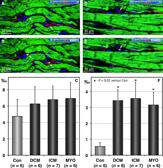
Proliferation and apoptosis of telocytes (TCs). (A and B) Confocal images showing typical TCs (red) in a patient with chronic myocarditis and heart failure. Arrows indicate 2 TCs that are Ki67-positive. Cardiomyocytes are stained green with phalloidin and nuclei are stained blue with 4,6-diamidino-2-phenylindole (DAPI). (C) The mean values of Ki67-positive TCs expressed as absolute numbers of such cells per 1000 TCs. (D and E) Confocal images showing typical TCs (red) being TUNEL-positive (arrow). Cardiomyocytes are stained green with phalloidin and nuclei are stained blue with DAPI. (F) Quantification of TUNEL-positive TCs.
