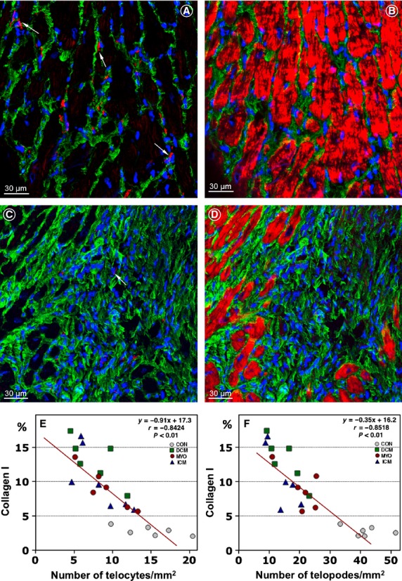
Figure 4.

Telocytes (TCs) are decreased in areas of collagen accumulations. Expression of collagen I (green) and the number and distribution of TCs (red) in a control patient (A and B) and in a patient with ICM (C and D). Note that TCs and telopodes (Tps) are scarce or absent in the area of replacement fibrosis which contains densely packed collagen fibres (C and D). Cardiomyocytes are stained red with Alexa633-phalloidin. Nuclei are stained blue with 4,6-diamidino-2-phenylindole (DAPI). (E and F) Correlations between the percent of collagen I per myocardial area and the number of TCs and Tps.
