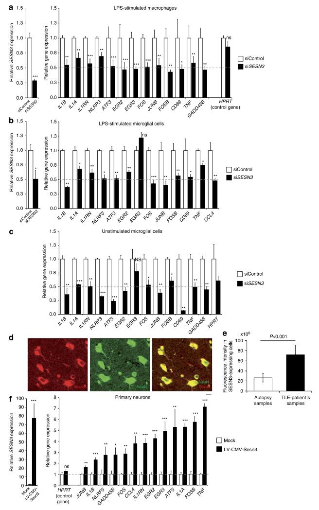
Figure 4. SESN3 regulates expression of Module-1 genes in macrophages, microglial cells and neurons.
Effect of siRNA-mediated knockdown of Sesn3 as compared with control siRNA (siControl), showing significant inhibition of Sesn3 mRNA expression and downregulation of Module-1 genes in murine LPS-stimulated (1 h) BMDM (a) and BV2 microglial cells (b), as well as in unstimulated BV2 microglial cells (c). Five independent biological replicates were used for BMDM experiments and at least three replicates in the BV2 microglia cells experiments. Data normalized to β-actin levels are shown as means relative to control ±s.e.m. (d) SESN3 immunofluorescence of human hippocampal slices from TLE patients: co-immunostainings with NeuN (green) antibody showed that SESN3 (red) is localized in neurons. Scale bar, 100 μm. (e) Quantification of SESN3 expression in human hippocampal tissue by immunofluorescence analysis. Maximum intensity projections of confocal z-stack images of immunohistochemical stainings with antibody against SESN3 were used. For determination of SESN3 cell fluorescence as a measure of SESN3 expression level, SESN3-expressing cells in the CA2 region of the hippocampus in both TLE patients samples (n = 7) and autopsy samples (n = 8) were measured using ImageJ software. Cell fluorescence was assessed as follows: integrated density—(area of selected cell × mean fluorescence of background readings). SESN3 total cell fluorescence in TLE patients is significantly increased as compared with the SESN3 total cell fluorescence in autopsy samples (two-tailed Mann–Whitney test, P<0.001). Fluorescence intensity data are reported as means±s.e.m. (f) Effect of lentiviral-mediated Sesn3 overexpression on Module-1 genes in primary hippocampal neurons. Left, relative levels of Sesn3 mRNA in transduced neurons (LV-CMV-Sesn3) compared with the levels in mock transduction (Mock). Right, relative mRNA levels of Module-1 genes and a control gene not in the network (Hprt) in transduced neurons compared with levels in mock transduction. Data normalized to Gapdh levels are shown as means relative to control ±s.e.m. Four (Mock group) and twelve (LV-CMV-Sesn3 group) replicates were used in neuronal cell experiments. Statistical significance of the differences (P-value) between siSESN3 (or LV-CMV-Sesn3) and siControl (or Mock) was assessed by t-test (two-tailed) and adjusting for unequal variances across different groups. *P<0.05; **P<0.01; ***P<0.001; ****P<0.0001; NS, not significant (P>0.05).

