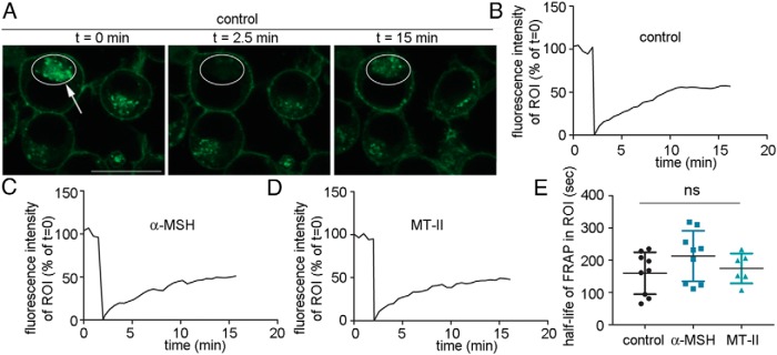
Figure 4.
HA-MC4R-GFP in a complex with MTII internalizes rapidly and at the same rate as the receptor in a complex with α-MSH or the ligand-free receptor. A, N2AHA-MC4R-GFP cells were transferred to a heated stage at 37°C for photobleaching/confocal microscopy. A, ROI is drawn around the HA-MC4R-GFP intracellular compartment of N2AHA-MC4R-GFP cells kept in basal conditions (t = 0, white circle). Photobleaching is carried out at the ROI (t = 2.5 min, white circle highlighted by the arrow). Fluorescence recovery is visualized at the ROI (t = 15 min, white circle). Scale bar, 10 μm. B–D, Integrated fluorescence intensity at the intracellular ROI is monitored over time in a cell either kept in basal conditions (A) or treated with α-MSH (C) and with MTII (D). The agonists, at a concentration of 200nM, were added to the cell medium 45 minutes before the start of the FRAP experiment. E, The graph shows the average half-life of FRAP in the selected ROIs (data are derived from ∼6 cells per condition).

