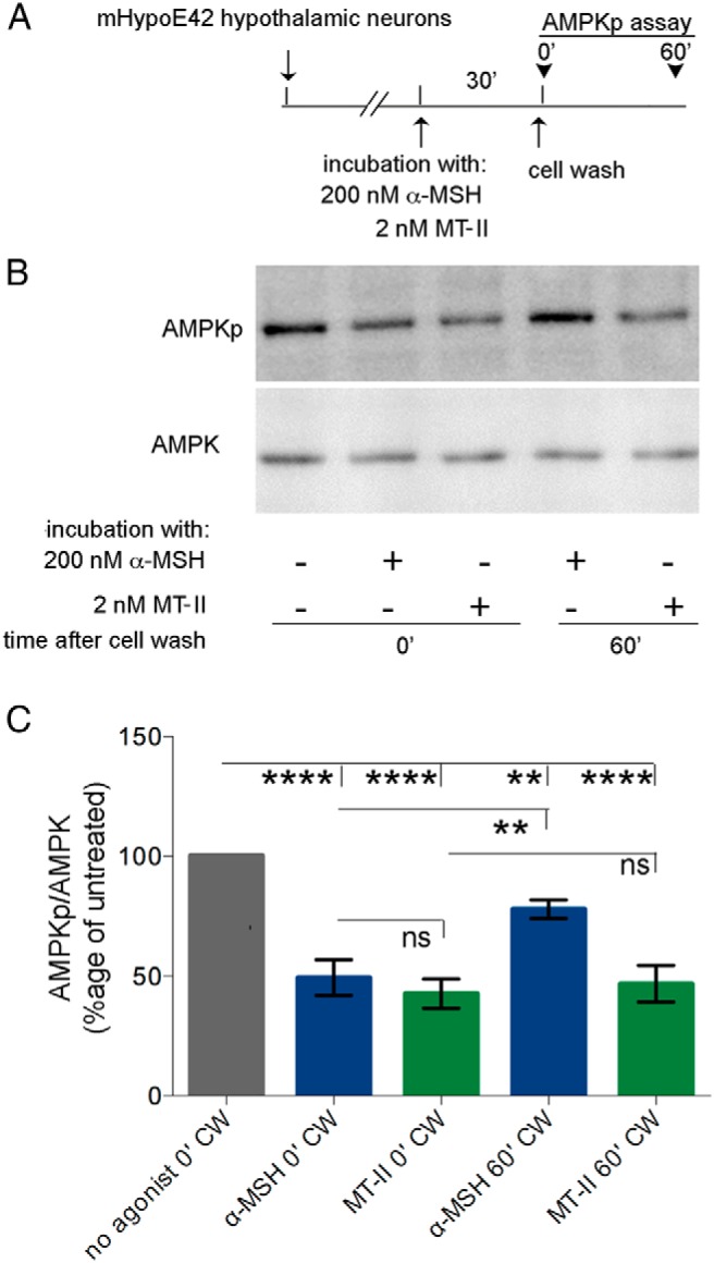
Figure 6.

MTII induces persistent AMPK signal in mHypoE-42 hypothalamic neurons. A, mHypoE-42 hypothalamic cells were treated as indicated by the schematic diagram. B, Western blot analysis of cleared mHypoE-42 hypothalamic neurons lysates with the indicated antibodies. C, The intensity of the AMPKp and AMPK bands were measured as described in Materials and Methods, and data were expressed as the percentage of the untreated control. Averages and SDs were derived from 3 independent experiments.
