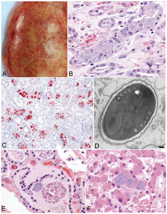
Figure 2.
Gross and microscopic pathology of tissues from the left kidney recipient.
A: Explanted kidney showing multiple microabscesses and hemorrhages of the renal capsule. B: Intracellular masses of Encephalitozoon cuniculi within renal tubular epithelium in the explanted kidney (original magnification × 100). C: Immunohistochemical staining of Encephalitozoon cuniculi (red) in the explanted kidney (original magnification × 25). D: Electron micrograph of a spore of Encephalitozoon cuniculi, surrounded by a dense exospore and electron-lucent endospore and containing 5 cross-sections through the coils of the polar filament, as is typical for this microsporidian (bar, 100 nm). E and F: Evidence of disseminated microsporidiosis involving thyroid gland (E) and liver (F) (original magnification × 100). Encephalitozoon cuniculi organisms were detected in several other tissues, including the central nervous system, heart, trachea, lung, pancreas, adrenal gland, gallbladder, prostate gland, stomach, colon, and vascular smooth muscle.

