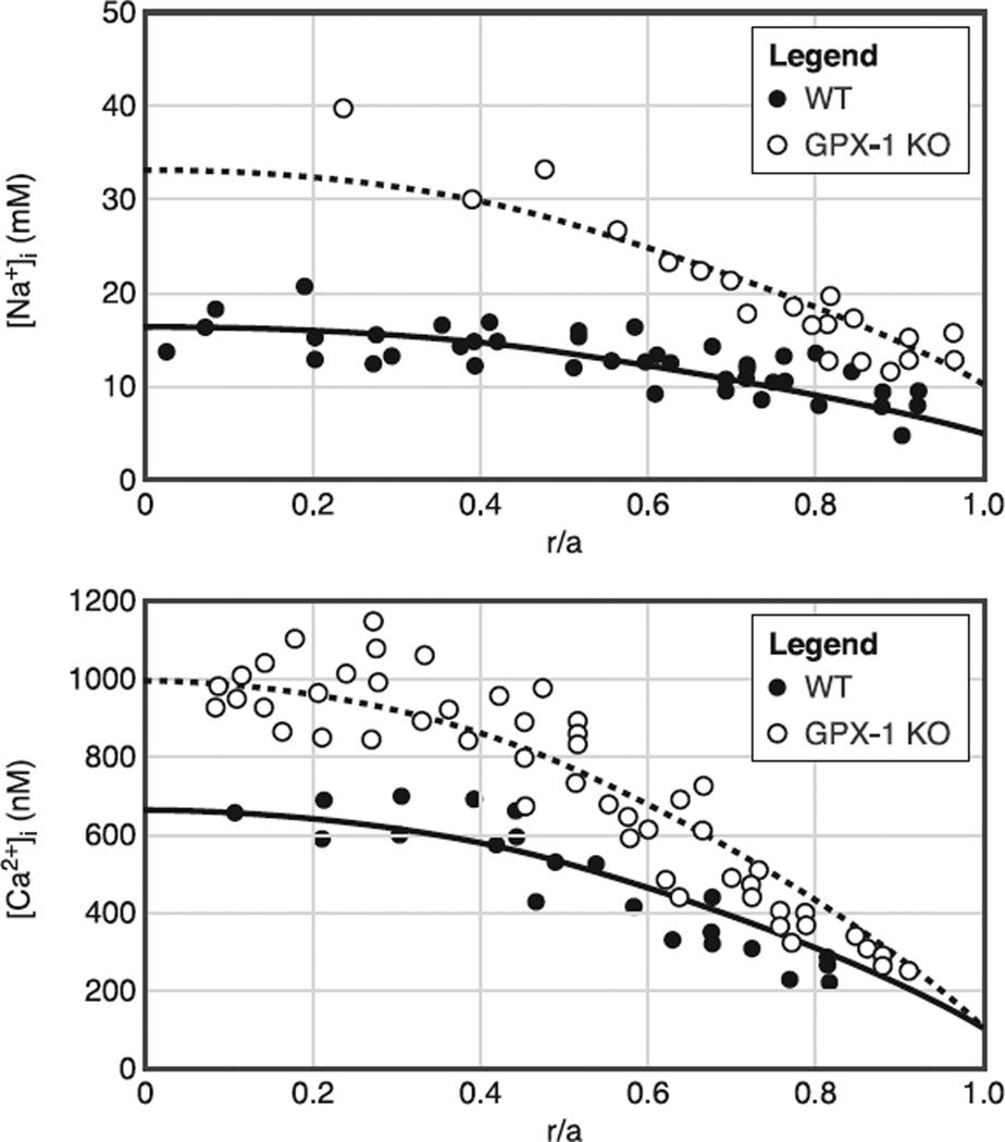
Fig. 15.
The effect on intracellular homeostasis of knocking out GPX-1, an enzyme that protects the lens against H2O2-mediated oxidative damage. All lenses were ~2 mo old. The smooth curves are from a structurally based model of the lens circulation. The only detectable defect in the GPX-1 KO lenses was that MF coupling conductance was on average half the value in WT lenses, so the expected effect is that the same circulation of Na+ or Ca2+ will require twice the center-to-surface concentration gradient. A: intracellular Na+ concentration gradients measured with SBFI in WT and GPX-1 KO lenses. In WT lenses, the Na+ concentration gradient is ~10 mM, as the concentration goes from 17 mM at the center to 7 mM at the surface. In the GPX-1 KO lenses, the gradient is ~22 mM, as the concentration goes from 33 mM at the center to 11 mM at the surface. B: intracellular Ca2+ concentration gradients measured with fura 2 in WT and GPX-1 KO lenses. In the WT lens, the gradient is ~500 nM, as the concentration goes from 650 nM in the center to 150 nM at the surface. In the GPX-1 KO lenses, the gradient is ~850 nM, as the concentration goes from ~1,000 nM at the center to 150 nM at the surface. [From Wang et al. (204), with permission from Springer Science + Business Media.]

