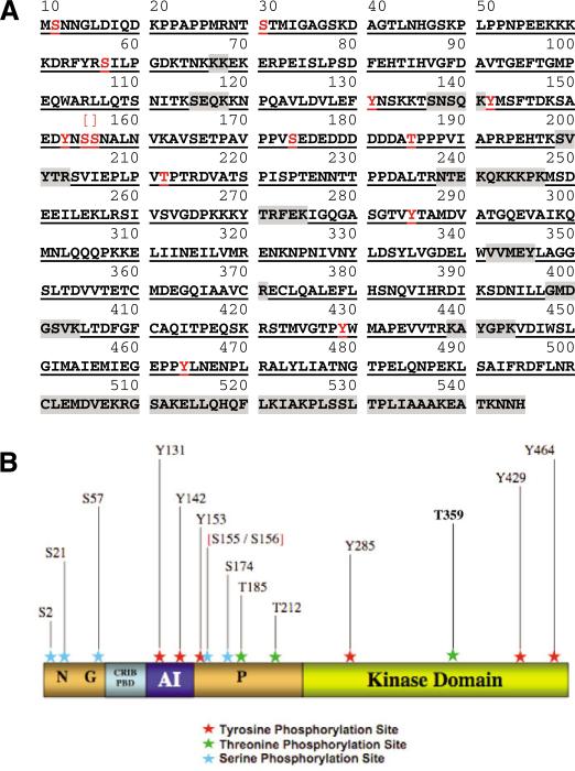
Fig. 2.
Phosphorylation sites detected in human PAK1. (A) Ser, Thr and Tyr coverage of the FLAG-PAK1 sequence (tag not shown) generated with trypsin/chymotrypsin. Detected peptides are bold and underlined. Residues not covered are shaded in gray. Observed phosphorylation sites are red. In total, 83% of the amino acid sequence was covered. Thirteen phosphorylation sites were identified. (B) Mapping of domains and identified phosphorylation sites. MS/MS-identified phosphorylation sites are labeled. N, G and P denote the NCK-, GRB2- and βPIX-binding sites, respectively; AI, auto-inhibitory domain. Red brackets indicate the ambiguous site.

