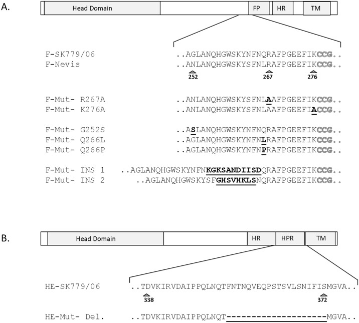
Fig 1. Schematic illustration of the structure of ISAV surface glycoproteins.
The F protein contains the predicted transmembrane domain (TM), heptad repeat (HR) and fusion peptide (FP) which is also highlighted in grey in the sequence (A). The HE contains a Highly Polymorphic Region (HPR) (B). HRs positions were predicted using the program LearnCoil-VMF (http://cis.poly.edu/~jps/matcher.html) (data no shown). Arrows indicate important aa positions in the proteins, while substitutions and insertions introduced into the mutant F proteins (A) and the deletion in the mutant HE (B) are in bold and underlined.

