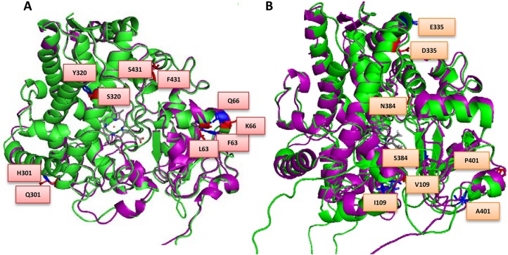
Fig 2. Overlay of (A) MALCYP6P9a (green helices) and FANGCYP6P9a (purple helices) and (B) MALCYP6P9b (green helices) and FANGCYP6P9b (purple helices) showing amino acid residues changes.
Key residues are in stick format and those belonging to MALCYP6P9a or MALCYP6P9b are highlighted in red and annotated, while corresponding residues in FANGCYP6P9a and FANGCYP6P9b are in blue colour. Heme atoms are shown in stick format and grey.

