
Fig 6. Human Brain Images.
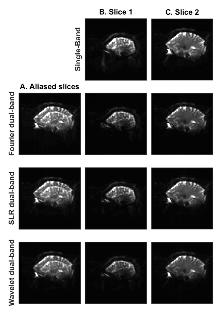
Reconstructed sagittal brain images obtained with the standard single-band pulse sequence on the General Electric MR950 7.0T scanner are compared to images obtained with the dual-band Fourier, SLR and wavelet-optimized refocusing pulses. All images are shown with the same window level. Single-band images are shown in the top row with standard reconstruction on the scanner. Column A shows the acquired (aliased) images containing two different brain slices superimposed. The two separated slices, after SENSE reconstruction, are shown in column B and column C for the Fourier, SLR and wavelet-optimized refocusing pulses. Other than differing refocusing pulses, all aspects of the scan sequence remained the same.