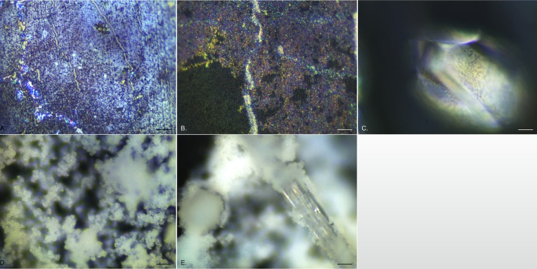
Fig 3. Representative EDIC images showing crystalline biofilm development on all silicone catheter sections exposed to P. mirabilis.
a. 24 h exposure showing multi-layered appearance and highly reflective, motile P. mirabilis. b. 3 days exposure showing development of a microcrystalline layer attaching to the initial conditioning film below. c. an individual struvite crystal formed after 3 days exposure. d. after 4 days exposure, copious amounts of diffuse crystalline material (apatite) formed, creating a thick three-dimensional structure. e. at the same time (and over the remaining time course), large, rod-shaped crystal embedded in diffuse crystalline material formed, extending in length to 10 mm. (Magnification a—d x 1000, bar = 10 μm; e x 500, bar = 20 μm).

