
Fig 3. Immunohistology of kidneys showing the cellular location of AQP2.
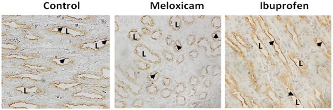
Perfusion fixed kidneys from control, meloxicam- and ibuprofen-treated mice were prepared for histology examination as described in the Methods section. AQP2 is denoted by the brown stain indicating positive peroxidase product. Slides were counterstained with hematoxylin to show nuclei. L indicates examples of the lumen of the collecting ducts. Arrows show the apical staining location of the AQP2. Magnification, 400x.