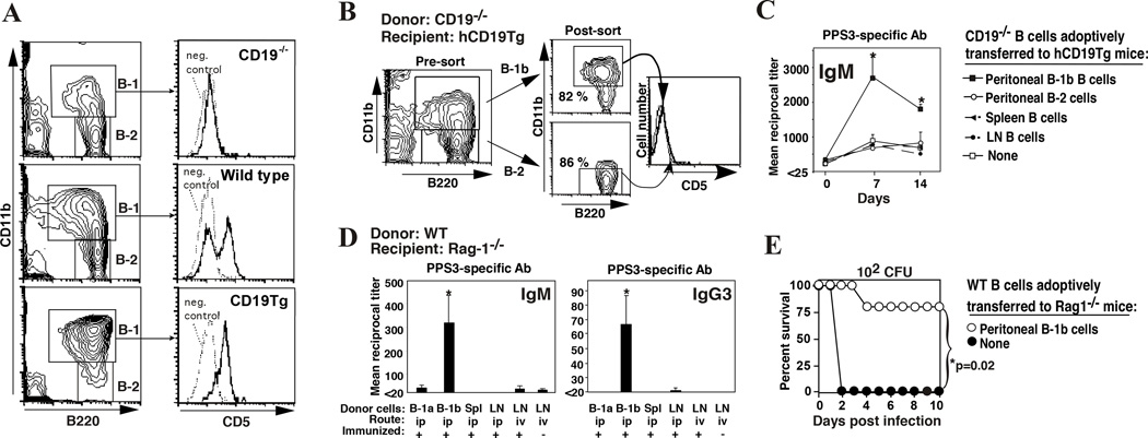
Figure 1. B-1b cells reconstitute protective antibody responses to PPS in B-1b-cell deficient CD19Tg mice and B cell-deficient Rag-1−/− mice.
A) CD19−/− mice are deficient in B-1a cells whereas CD19Tg mice are deficient in B-1b cells. B-1 (B220+CD11b+) and B-2 (B220+CD11b−) lymphocytes are indicated (left column) with histograms showing CD5 expression by peritoneal B-1 (B220+CD11b+-gated) cells (right column). Isotype-matched control antibody staining is indicated by a dotted line. B–C) Reconstituting hCD19Tg mice with peritoneal B-1b cells from CD19−/− mice rescues responsiveness to PPS-3. Peritoneal B-1b or B-2 cells from CD19−/− mice were isolated by FACS (B). FACS-purified peritoneal B cells or enriched spleen and lymph node B cells from CD19−/− mice were transferred i.p. into hCD19Tg mice (105 cells/mouse). Mice were immunized with PPS-3 3 weeks later with PPS-3-specific antibody titers determined by ELISA (C). D–E) Transfer of WT B-1b cells into Rag-1−/− mice reconstitutes PPS3-specific IgM and IgG responses and provides protection against lethal S. pneumoniae infection. D) Purified WT peritoneal B-1a cells, B-1b cells, or unfractionated spleen or LN cells were transferred i.p. or i.v. into Rag-1−/− mice (4 × 10⁵ B cells/mouse; ≥3 mice/group). Mice were immunized with 0.5 µg PPS-3 3 days later, with PPS-3-specific IgM (d7) and IgG3 (d14) antibody levels measured by ELISA. E) Rag-1−/− mice reconstituted with B-1b cells were infected with 102 colony forming units of serotype 3 S. pneumoniae 14 days post-immunization. *Chi-square analysis indicated significant differences in survival. Adapted from Haas et al.8.

