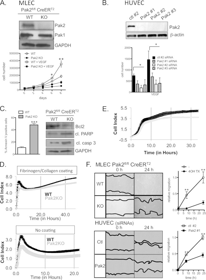
FIG 3.
Pak2 KO and KD inhibit proliferation, attachment, and migration of ECs and enhance apoptosis. (A) Proliferation of MLECs in the presence and absence of VEGF. Western blot analysis of Pak1 and Pak2 expression in CreERT2; Pak2fl/fl MLECs treated with vehicle and 4-OHTx. GAPDH, glyceraldehyde-3-phosphate dehydrogenase. (B) Proliferation of HUVECs transfected with control (ctl) short hairpin RNA (shRNA) or three different Pak2 shRNAs. The results of Western blot analysis of Pak2 expression are shown. (C) Apoptosis analysis of CreERT2; Pak2fl/fl MLECs treated with vehicle and 4-OHTx. (Left) Percentage of annexin V-positive MLECs. (Right) Western blot analysis of apoptosis markers Bcl2, cleaved procyclic acidic repetitive protein (PARP), and cleaved caspase 3 from MLEC lysates. (D) Long-term cell attachment assay with MLECs on fibronectin/collagen-coated or noncoated plates. (E) Chemotactic migration of MLECs toward 10% serum. (F) Wound healing assay using MLECs and HUVECs. Images show the initial wound width and wound closure after 24 h. Relative wound migration was quantified from images at 0, 8, and 24 h. Error bars represent SD in all panels. Experiments were repeated more than 4 times. Symbols: *, P < 0.05; **, P < 0.01; ***, P < 0.001.

