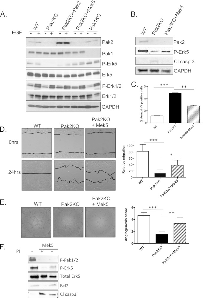
FIG 7.
Pak2 regulates the Mek5/Erk5 signaling pathway. (A) Western blot analysis of MLEC protein lysates. MLECs were infected with adenovirus expressing WT-Pak2 and CA-Mek5 for 24 h and then treated with 4-OHTx. The cells were serum starved for 24 h and then induced with 10 ng/ml EGF (+) for 10 min. (B) Western blot analysis of apoptosis markers in protein lysates from MLEC cells infected with CA-Mek5. (C) Apoptosis assay showing percent annexin V-positive MLECs. (D) Wound healing assay with WT, Pak2 KO, and CA-Mek5 reconstituted MLECs. Error bars represent SD. (E) Tubule network assay with WT, Pak2 KO, and CA-Mek5 reconstituted MLECs. At least five different fields were given an angiogenesis score according to the manufacturer's recommendation. Error bars represent SD. Symbols: *, P < 0.05; **, P < 0.01; ***, P < 0.001. (F) WT MLECs and MLECs overexpressing constitutively active Mek5 were treated with Frax1036 pan-PakI inhibitor (PI) for 6 h. Western blot analysis of P-Erk5 and apoptosis markers shows that inhibition of Pak1 to Pak3 results in decreased active Erk5 and that Mek5 can partially rescue apoptosis events due to Pak2 loss.

