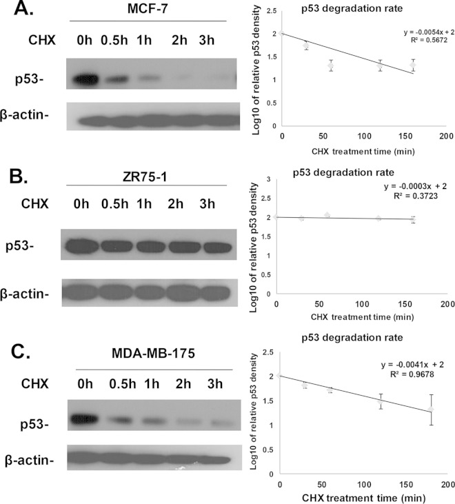
FIG 4.
Comparison of the p53 degradation rates in MCF-7, ZR75-1, and MDA-MB-175 cells. Confluent MCF-7 (A), ZR75-1(B), and MDA-MB-175 (C) cells were either left untreated or treated with 50 μg of cycloheximide/ml for 30 min, 1 h, 2 h, or 3 h. The cells were collected and lysed at each time point. Cell lysates were then subjected to SDS-PAGE and transferred onto a PVDF membrane. Immunoblotting to detect p53 was performed using a primary antibody against p53 and an HRP-conjugated secondary antibody for enhanced p53 signal. The levels of β-actin were also detected by Western blotting. The results shown (left panels) are representative of three experiments. Calculation of the p53 degradation rates from three individual experiments performed in different cell lines (right panels) was as described in Materials and Methods. The results indicate that the p53 half-lives were 56, 1,003, and 74 min for MCF-7, ZR75-1, and MDA-MB-175 cells, respectively.

