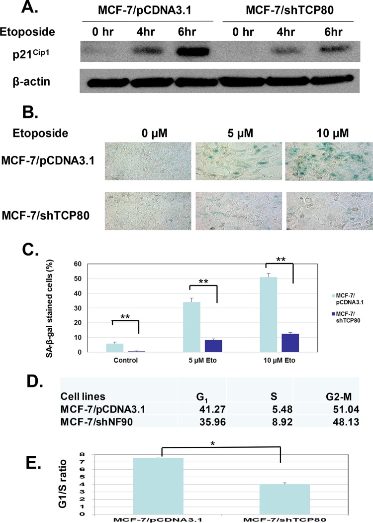
FIG 8.
MCF-7/shTCP80 cells exhibit diminished ability to induce G1 cell cycle arrest and senescence following DNA damage compared to MCF-7/pCDNA3.1 cells. (A) p21Cip1 induction following DNA damage is decreased in MCF-7/shTCP80 cells. Subconfluent MCF-7/pCDNA3.1 and MCF-7/shTCP80 cells were treated with 10 μM etoposide for 4 or 6 h. After treatment, the cells were lysed, and equal amounts of protein were subjected to SDS-PAGE and Western blotting. p21Cip1 and β-actin proteins were detected with their respective antibodies. The results presented are representative of three separate experiments. (B and C) MCF-7/shTCP80 cells exhibit diminished ability to induce senescence following etoposide treatment. Subconfluent MCF-7/pCDNA3.1 and MCF-7/shTCP80 cells were treated with 5 or 10 μM etoposide for 4 days. After treatment, the medium was removed, and the cells were washed with PBS. The cells were then stained for SA-β-galactosidase using a kit from Cell Signaling as described in Materials and Methods, and pictures were taken with a microscope (Leica DM IRB) at ×40 magnification. The number of SA-β-Gal-positive cells versus total cells per field in three independent fields of view was counted. The results in panel C represent the means ± the SEM of three independent measurements (**, P < 0.01). (D and E) Etoposide causes stronger G1 cell cycle arrest in MCF-7/pCDNA3.1 cells. Subconfluent MCF-7/pCDNA3.1 and MCF-7/shTCP80 cells were treated with 10 μM etoposide for 24 h. After treatment, cells were collected and subjected to flow cytometry analysis after being fixed with ethanol and stained with propidium iodide. The cell population at each phase was analyzed by the Modfit 2 software. The G1/S ratio presented in panel E represents the average ± the SEM from three individual experiments (*, P < 0.05).

