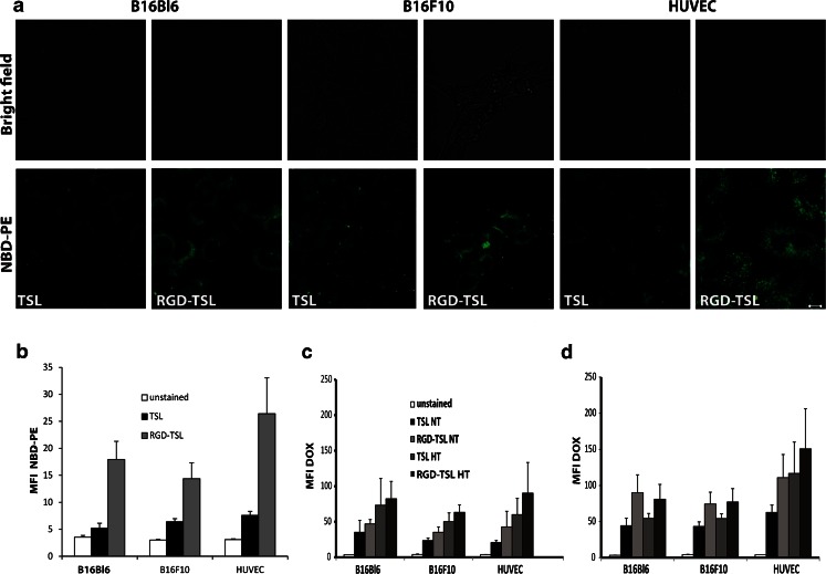
Fig. 3.
Confocal live-cell imaging for preferential uptake of RGD-TSL compared to TSL into B16Bl6, B16F10 and HUVEC cells after 3 h of incubation at 37°C (a). Unbound liposomes were removed by washing. Scale bar applies for all images, 10 μm. Intracellular NBD-PE fluorescent intensity represented as mean fluorescent intensity (MFI) in melanoma B16Bl6, B16F10 cells and HUVEC (b) treated with either TSL or RGD-TSL for 3 h at 37°C. Unbound liposomes were removed by washing. As unstained cells are used cells which were not incubated with liposomes. c and d. Intracellular Dox uptake represented as MFI in melanoma B16Bl6, B16F10 cells and HUVEC after 1 h (c) or 3 h (d) of incubation at 37°C, washing of unbound liposomes, followed by 1 h of HT at 42°C.

