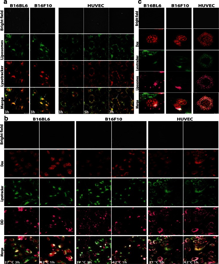
Fig. 4.
(a). Confocal microscopy on melanoma B16Bl6 and B16F10 cells and HUVEC incubated with NBD-PE (green) labelled RGD-TSL for 3 h at 37°C and lysotracker (red). Unbound liposomes were removed by washing 3× with medium without FCS. After washing, B16Bl6 and B16F10 were immediately imaged at 37°C, whereas HUVEC were followed up to 7 h. Internalized liposomes in the cytosol can be observed in all the cell lines (white arrows). Non-internalized in the lysosomes liposomes were also visible (blue arrows). B16Bl6 and B16 internalized liposomes immediately in the lysosomes after the 3 h of incubation period (yellow colocalization of green liposomes and red lysotracker), whereas this process happened in HUVEC after 7 h. Images were taken by confocal microscope (40×, 2,5 μm pinhole, 2× zoom). Scale bar applies for all images, 20 μm. (b). Doxorubicin release (red) from DiD-labelled RGD-TSL (purple) in B16Bl6, B16F10 and HUVEC upon HT trigger. Cells were incubated with 50 μM Dox for 3 h at 37°C, after which cells were washed 3× with medium without FCS. Images were taken right after this incubation at 37°C. Then, HT at 42°C for 1 h was applied and images in the end of the HT treatment were recorded. Images were taken by confocal microscope. Scale bar applies for all images, 50 μm. (c). Colocalization of RGD-TSL (purple) with lysotracker (green) and Dox release (red) in the lysosomes. Scale bar applies for all images, 10 μm.

