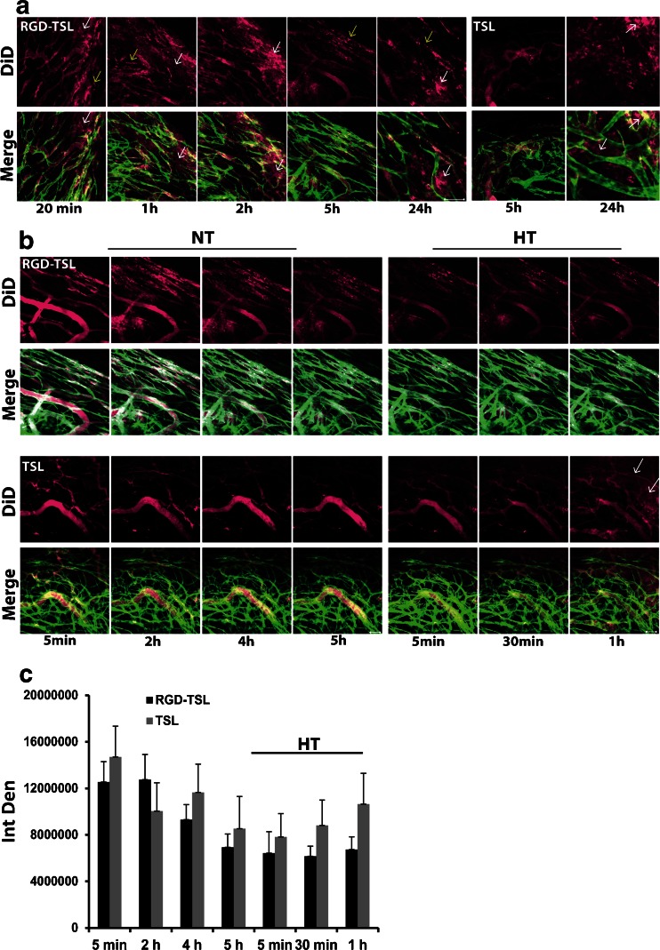
Fig. 6.
(a). Binding of DiD-labeled RGD-TSL (purple) to tumor vasculature (green) of B16Bl6 window chamber bearing mice. Binding of liposomes to tumor endothelial cells started 20 min after injection and was followed in time up to 24 h. Representative images from intravital microscopy were selected. Scale bar applies to all images, 50 μm. (b). RGD-TSL and TSL appearance (DiD, in purple) in tumor vasculature (green) during 5 h at NT in B16Bl6 window bearing mice (Fig B left panels) and upon subsequent HT at 42°C for 1 h (Fig. 5b right panels). DiD-labelled RGD-TSL or TSL were injected i.v, after which they were allowed to circulate in blood stream at NT for 5 h in order to allow binding of RGD-TSL to angiogenic endothelial cells. Thereafter, HT at 42°C for 1 h was applied to promote extravasation of RGD-TSL and TSL. Scale bar applies to all images 50 μm. (c). In vivo quantification of DiD liposomal fluorescence before and during 1 h of HT, presented as integrated density (IntDen) in time, see Materials and methods for details.

