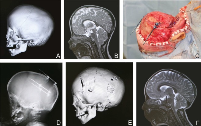
Fig. 3.
A 1 year and 10 months old male child with multiple suture synostosis. A: Preoperative 3D-CT. Sagittal and bilateral lambdoid sutures were fused. B: Preoperative MRI image. Chiari malformation was noted. C: Intraoperative view. D: Skull X-P just after 32 mm distraction. E: 3D-CT at 1 year and 3 months after frontal remodeling. F: MRI image at 1 year and 3 months after frontal remodeling. Reduction of Chiari malformation was demonstrated. 3D-CT: three dimensional computed tomography, MRI: magnetic resonance imaging.

