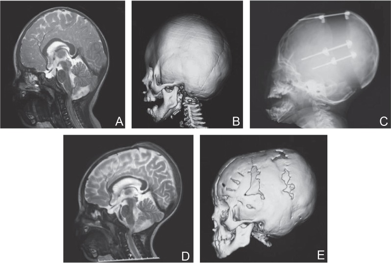
Fig. 4.
A 1 year and 3 months old boy with Saetre-Chotzen syndrome. A: Preoperative MRI. Significant stenosis of posterior in cerebellum and medulla oblongata region was seen. B: Preoperative 3D-CT image. C: Plain X-P after completion of 30 mm distraction. D: MRI image after distraction. Posterior stenosis was released and subarachnoid space was expanded. Note the change of the shape of corpus callosum. E: 3D-CT image at 1 year after fronto-orbital remodeling. 3D-CT: three dimensional computed tomography, MRI: magnetic resonance imaging.

