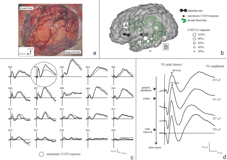
Fig. 2.
Cortico-cortical evoked potential (CCEP). a: Intraoperative view of electrodes placement over frontal/temporal lobe, arranged with tumor lesion. b: Scheme of electrodes placement and CCEP distribution under general anesthesia. Electrical stimuli were delivered on electrodes A02 and A07. CCEP distributed maximum at electrode B02 over the middle to posterior part of the temporal lobe. c: CCEP waveforms in plate B, before tumor removal, under awake condition. Two trials were superimposed to check the reproducibility. CCEP distribution did not change under between general anesthesia and awake condition. d: CCEP change along with surgical procedures at the maximum CCEP response site (N1 amplitude in electrode B02). CCEP waveforms were sequentially shown from the top to the bottom along the time course. As the patient became awake, the N1 amplitude increased from 215 mV to 311 mV (145%). After tumor removal the N1 amplitude did not decline (329 mV). The patient preserved normal language function throughout the perioperative period. Adapted with permission from Reference 50.

