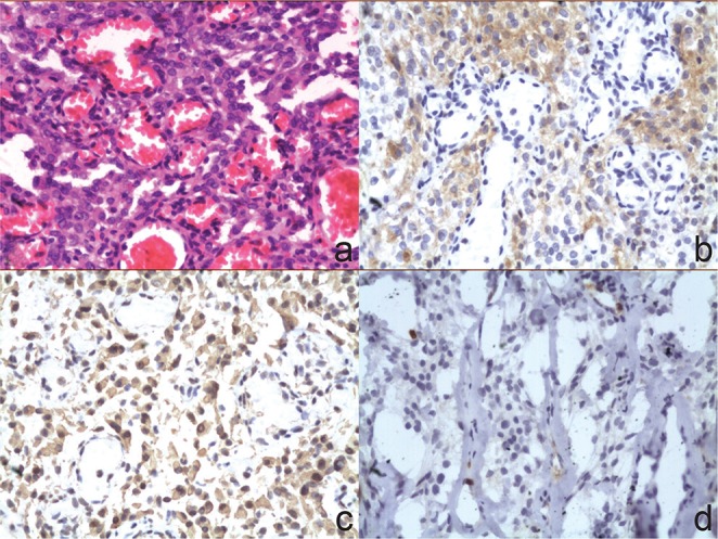
Fig. 5.

Photomicrographs illustrated that the tumor consisted of dilated vascular spaces with intervening areas showing spindle to oval cells with abundant cytoplasm and oval nuclei. There was no evidence of cytonuclear atypia, necrosis, or mitotic activity (a: H&E stain, original magnification ×200). The tumor cells were positive for epithelial membrane antigen (b: Immunohistochemical stain, original magnification ×200), vimentin (c: Immunohistochemical stain, original magnification ×200). The Ki67 proliferation index was 1.9% (d: Immunohistochemical stain, original magnification ×200). H&E: hematoxylin and eosin.
