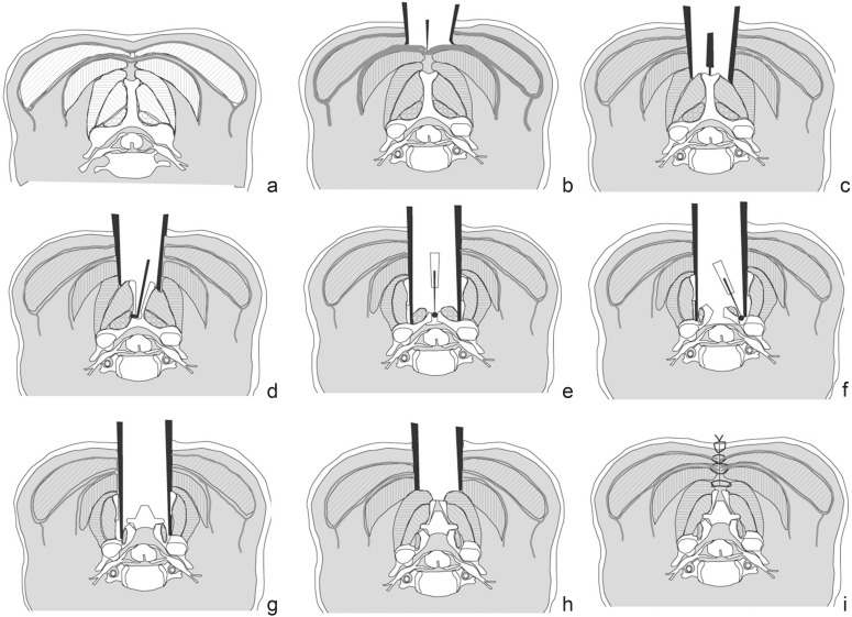
Fig. 5.
Schematic drawing depicting Kim’s myoarchitectonic spinolaminoplasty, in which attachments of all posterior muscles are preserved while expanding the spinal canal. Cross section of the cervical spine and layers of nuchal muscles: the trapezius in the 1st layer, the splenius in the 2nd layer, and the semispinalis capitis in the 3rd layer of the nuchal ligament. a: Semispinalis cervicis and multifidus are attached to the sides of the spinous process. b: The fascia in the midline of the trapezius is sharply cut. c: The midline fascia of the splenius is sharply divided and the tip of the spinous process is proved. d: The spinous process is split in the midline with a sagittal saw, and through this opening the spinous process is cut off from the lamina. e: The spinous process is retracted laterally with the semispinalis cervicis attachment intact. The lamina is drilled in the midline. f: Hinge gutters are made at the border between the lamina and the lateral mass, preserving the attachment of the multifidus. g: The laminal flaps are elevated on the hinges and are inter-bridged using a hydroxyapatite implant. h: The split halves of the spinous process with the attached semispinalis cervicis is attached to the hydroxyapatite implant. i: The Three layers of the nuchal ligament is firmly reconstructed in the midline.

