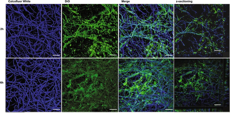
Fig. 3.

Confocal laser scanning microscopy images of two-day-old biofilms of A. fumigatus strain Af293. The fungal biofilms were stained with calcofluor white (blue staining). SLNs labelled with DiO (green staining) were then added and the penetrance measured after 2 h (upper panels) and 6 h (lower panels). The rectangular micrographs on the sides (right panels) represent the x–z plane and y–z optical cross sections through the thickness of the biofilms. The images shown (CLSM 20X) are representative of three independent experiments. Bar = 50 μm
