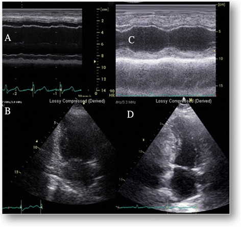
Fig. 1.

Echocardiographic findings on admission and early follow-up. a Admission, Basal MMode showing dilated left ventricle with severe systolic function. b Admission, end systolic apical 2-chambers view demonstrating large end systolic volume. c Follow-up at 3 weeks, Basal MMode showing normal ventricular size and contraction. d Follow-up at 3 weeks, end systolic apical 2-chambers view with normal ventricular size and contraction
