
Fig. 2.
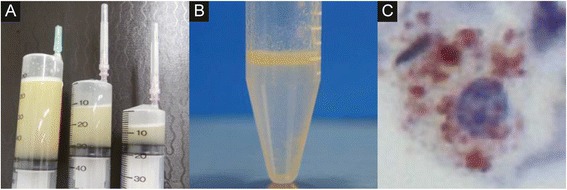
The bronchoalveolar fluid examination in a 66-year-old male with lipoid pneumonia. The bronchoalveolar lavage fluid was initially turbid white (a) and subsequently exhibited a bilayer appearance (b). Oil phagocytosis by alveolar macrophages was observed under a microscope with Sudan III staining (c)