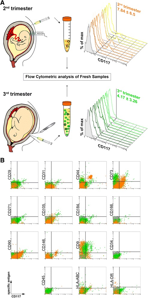
Fig. 1.

Cell isolation from collected amniotic fluid (from the second and third trimesters) and characterization by flow cytometry analysis. a Representative scheme of amniotic fluid retrieval for cell extraction from second-trimester amniocentesis (upper part) and cesarean section at term of pregnancy (below). Flow cytometry phenotyping revealed the variable presence of CD117 marker on freshly isolated cells (three-dimensional histograms, n = 10, isotype control in gray). b Representative dot-plot cytograms showing cells from both trimesters (second trimester in orange and third trimester in green) comparing various antigens (vertical axes) versus CD117 expression (horizontal axes)
