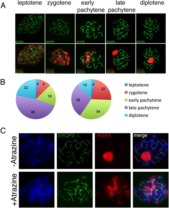
Fig. 2.

Atrazine causes delayed meiosis in mice. a γH2AX staining changes during meiotic prophase I substages. b The distribution of meiotic substages in control (left panel) and ATZ-treated (right panel) mice; 600 cells from control and treated mice were classified according to γH2AX and SYCP3 staining. Note that the number of spermatocytes in the zygotene and early pachytene stages is increased in ATZ-treated animals, while the number of spermatocytes in the late stages decreased. c Surface spreads from control and ATZ-treated testis stained with anti-γH2AX (red) and anti-SYCP3 (green) antibodies. In control mice, the majority of meiotic cells have γH2AX marks at the sex bodies only. In ATZ-treated mice, the majority of meiotic cells have γH2AX marks at many chromosomes
