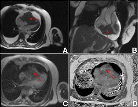
Fig. 2.

Preoperative patient 2 Heart MRI. a 4 chambers GRE view. Hypointensive masses visible in right ventricle, right atrium and right atrial appendage (red arrow). The tumour is obstructing the right ventricle and compressing the interventricular septum, and the left ventricle. b 2 chambers GRE view. Hypointensive masses in the right ventricle and right atrium (red arrow). c 4 chambers view perfusion sequence. Perfusion is seen in the masses (red arrow). d 4 chambers view. Non homogenous gadolinium enhancement is visible (red arrow). All images were taken during diastole
