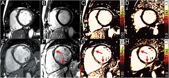
Fig. 4.

Two patients with non-ischemic dilated cardiomyopathy. (a, e) Dilation of the left ventricle is present in both patients on end-diastolic images. In the first patient, (b) no LGE-enhancement is present, (c) ΔS/So is normal throughout the heart as is (d) native-T1. In the second patient, (f) mid-wall septal LGE-enhancement is present (red arrows). (g) Heightened ΔS/So is observed in close agreement with the LGE image (red arrows), however (h) native-T1 values are elevated primarily at the right ventricular insertion-point
