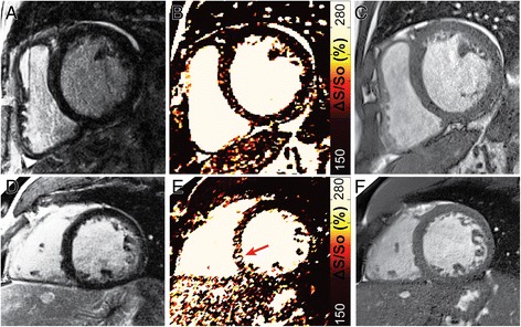
Fig. 8.

False positive identification of enhancement at ΔS/So. (a-c) Scattered noise on ΔS/So maps led to the false identification of diffuse enhancement in 3 of the 4 false positive cases. A representative example of a patient without enhancement at LGE (a) that was classified by blinded readers as demonstrating diffuse enhancement at ΔS/So (b) in the septum with corresponding anatomical reference image (c). d-f In one patient without enhancement at LGE (d), focal enhancement (arrow) was identified on the corresponding map of ΔS/So, with corresponding anatomical image shown in (f)
