Figure 3.

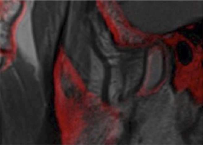
Sagittal image (MRI-CBCT marker-guided registration) of the right temporomandibular joint of Subject 5 showing imperfect overlap contours and edges of the condyle, mimicking mild motion artefact. Registration quality was ranked as fair.

Sagittal image (MRI-CBCT marker-guided registration) of the right temporomandibular joint of Subject 5 showing imperfect overlap contours and edges of the condyle, mimicking mild motion artefact. Registration quality was ranked as fair.