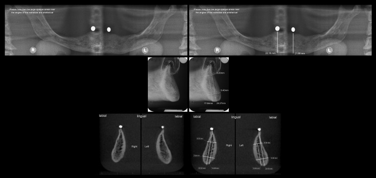
Figure 6.
A full set of images, for one of the four mandibles, presented to participants. Top row—panoramic radiographs without and with measurements; middle row—trans-symphyseal view without and with measurements; bottom row—CBCT cross-sectional images at the sites of implant placement without and with measurements. L, left; R, right.

