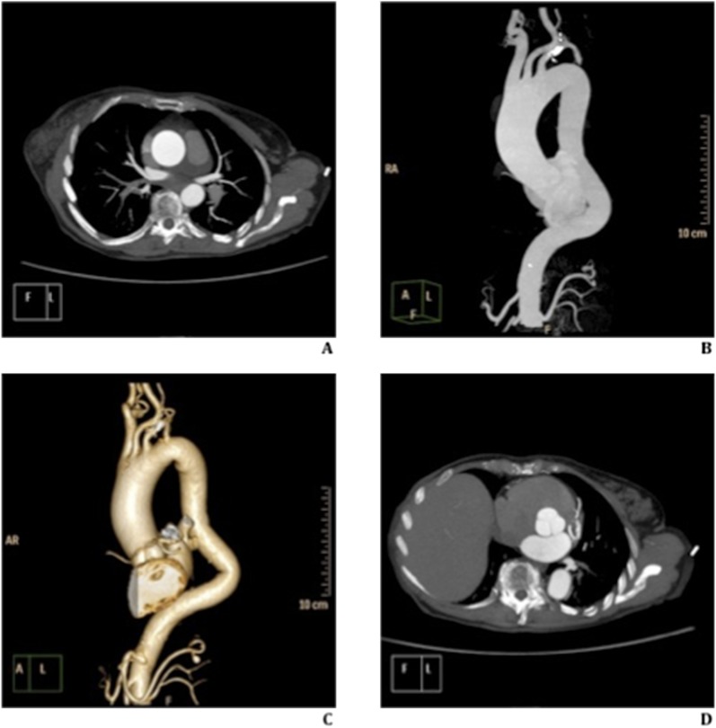
Figure 1.
73 year old-female with ascending aorta aneurysm. Thoracic aorta ECG-gated study, with low tube voltage (100 kV) and ultra low contrast material (40 ml). (a–d) Axial (a and d) and parasagittal (b) maximum intensity projections and volume-rendering reconstruction (c) show homogeneous and uniform contrast enhancement of vascular structures analysed. High definition of ECG-gated study, without cardiac motion artefacts, highlights the aortic valve (d).

