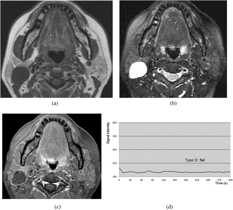
Figure 7.
Lymphoepithelial cyst of the right parotid gland (51-year-old female). (a) T1 weighted image reveals a hypointense mass (arrow). (b) T2 weighted image with fat suppression reveals a mass with homogeneous high signal intensity. (c) Post-contrast T1 weighted image with fat suppression reveals no enhancement of the mass except for the wall. (d) The time–signal intensity curve has Type D pattern.

