Figure 9.
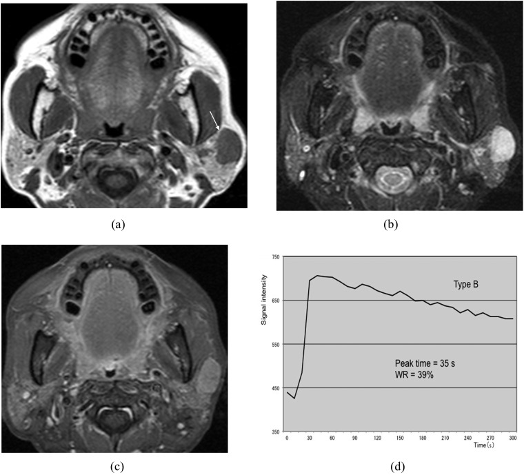
Malignant lymphoma of the left parotid gland (62-year-old female). (a) T1 weighted image reveals a hypointense mass with a relatively well-defined border (arrow). (b) T2 weighted image with fat suppression reveals a mass with homogeneous high signal intensity. (c) Post-contrast T1 weighted image with fat suppression reveals almost homogeneous tumour enhancement. (d) The time–signal intensity curve has Type B pattern [peak time of 35 s and washout ratio (WR) of 39%].

