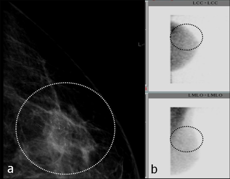
Figure 10.
(a, b) Small, breast-specific gamma imaging (BSGI)-negative ductal carcinoma in situ in the right upper outer quadrant. Dashed ellipses encircle microcalcifications. Ellipses in (b) mark the expected location of microcalcifications in BSGI. No Technetium-99m SestaMIBI uptake could be seen. (a) Mammogram [craniocaudal (cc)], magnification view; (b) BSGI in cc (figure above) and mediolateral-oblique (figure below) projection. LCC, left craniocaudal; LMLO, left mediolateral oblique.

