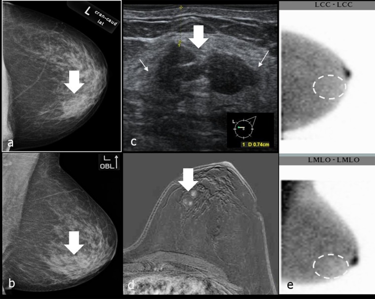
Figure 11.
(a–e) A breast-specific gamma imaging (BSGI) negative, breast imaging reporting and data system (BI-RADS®) IVa lesion (indistinct lateral margins in ultrasound) in the left breast. Histopathological examination confirmed fibroadenomatosis. Thick arrows in (a–d) point to the BI-RADS IVa lesion, thin arrows in (c) mark the indistinct margins of the lesion in ultrasound. Dashed ellipses in (e) mark the expected location of the lesion in scintigram. (a, b) Mammography (MGs) of the left breast craniocaudal (cc) (above) and mediolateral-oblique (mlo) (below) projection; (c) ultrasound of the BI-RADS® IVa lesion; (d) MRI of the left breast, T1wFS + DOTAREM® (Guerbet, Cedex, France); BSGIs of the left breast in cc (figure above) and mlo (figure below) projection. LCC, left craniocaudal; LMLO, left mediolateral oblique.

