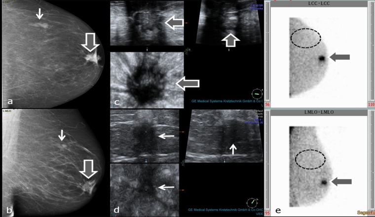
Figure 6.
(a–e) Characterization of mass lesions with breast-specific gamma imaging (BSGI). Thick arrows in (a–c) and (e) point to invasive ductal carcinoma behind the left nipple, thin arrows in (a, b and d) point to circumscribed breast parenchyma with fibrosis. Dashed ellipses in (e) encircle the region of upper outer quadrant without tracer uptake (assumed position of second lesion). (a, b) Mammograpy craniocaudal (cc) (a) and mediolateral oblique (mlo) (b) projections; (c) three-dimensional (3D) ultrasound of carcinoma behind the nipple; (d) 3D ultrasound of breast parenchyma with fibrosis; (e) planar BSGI in cc (figure above) and mlo (figure below) projections.

