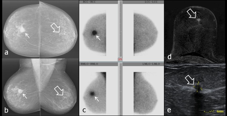
Figure 9.
(a–e) Breast-specific gamma imaging (BSGI)-occult, small left-sided carcinoma (G2). Left breast was assessed as Category 2 according to Brem et al.13 Larger right-sided carcinoma (18 mm) was clearly seen as focal spot on scintigram. Thick arrows in (a, b, d and e) point to 4-mm left-sided carcinoma. Thin arrows in (a–c) mark the 18-mm right-sided carcinoma. (a, b) Bilateral mammograms craniocaudal (cc) (above) and mediolateral oblique (mlo) (below); (c) BSGIs of both breasts in cc (figure above) and mlo (figure below) projections; (d) MRI of the left breast, T1 weighted FS + DOTAREM® (Guerbet, Cedex, France); (e) ultrasound of the left-sided cancer. LCC, left craniocaudal; LMLO, left mediolateral oblique; RCC, right craniocaudal; RMLO, right mediolateral oblique.

