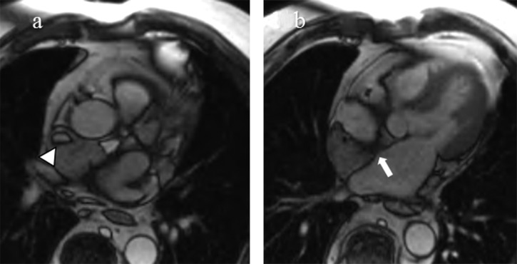
Figure 10.
(a, b) Lipomatous hypertrophy of the interatrial septum in a 63-year-old male with history of right atrial mass seen on echocardiography. Balanced steady-state free precession MR images demonstrate a large amount of tissue in the interatrial septum. This tissue follows signal characteristics of fat on all sequences with signal loss on fat saturated sequences (not shown). Note narrowing of the superior vena cava (arrowhead) and sparing of the fossa ovalis (arrow).

