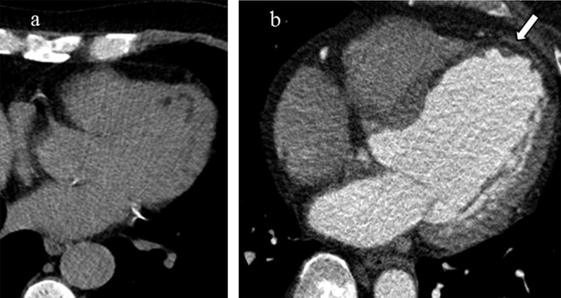
Figure 15.
(a, b) Lipomatous metaplasia in chronic myocardial infarct in two different patients. Transaxial non-contrast CT image of the heart demonstrates curvilinear low attenuation in a subendocardial distribution along the anterolateral and apical walls of the left ventricle. In a different patient, there is transmural fibrofatty replacement and thinning along the interventricular septum, anteroseptal wall and apex of the left ventricle (arrow) consistent with prior myocardial infarct in the left anterior descending distribution.

