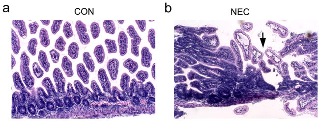
Figure 2.

Histopathology of small intestine sections (H&E staining) from control and NEC pups. (a) Representative control sample shows the normal villous structure with intact crypt region. (b) The induction of NEC led to the disruption of villous structure (arrow). Magnification: x200.
