Figure 3.
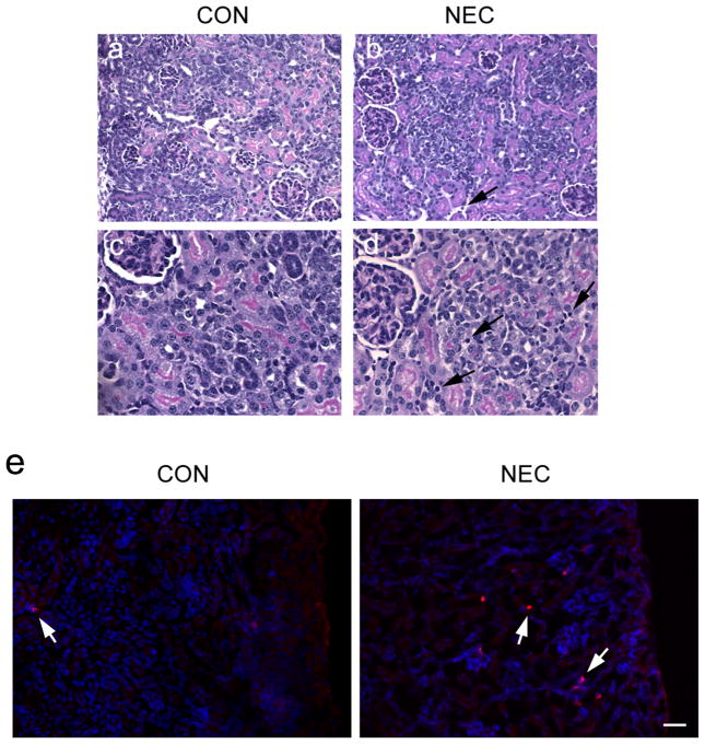
Histopathology of kidneys and the infiltration of immune cells. The control (a, c) and NEC (b, d) kidney sections from cortex regions (H&E staining) show normal glomerular and tubular architecture. However, NEC kidneys display the interstitial edema and mononuclear cell infiltration (arrows). (e) Frozen sections of control and NEC kidneys were stained with the CD11b antibody (red) recognizing monocytes and macrophages (arrows). The hoechst (blue) was used to stain nuclei. Scale bar: 60 μm.

