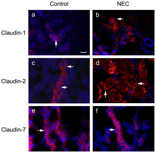
Figure 5.
Localization of claudin-1, -2 and -7 in control and NEC kidneys. Control and NEC kidneys were removed from the body and frozen in liquid nitrogen. Frozen sections (5 μm thickness) were immunostained with anti-claudin-1 (a, b), -2 (c, d) and -7 (e, f) antibodies. Arrows in a, b, c, e, and f indicate the conventional TJ staining and arrows in d point to the altered staining pattern. Scale bar: 20 μm.

