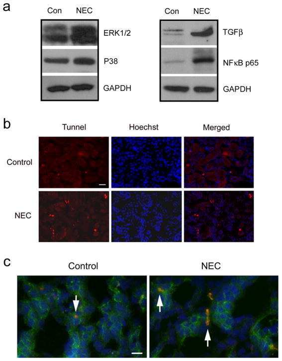
Figure 7.
Increased levels of inflammatory marker proteins and apoptotic cells in NEC kidneys. (a) Control and NEC kidney lysates were loaded on the SDS-polyacrylamide gel. Membranes were blotted against ERK1/2, p38, TGFβ and NFκB p65. GAPDH served as a loading control. (b) Control and NEC kidneys were fixed with 100% acetone and incubated with 10% BSA in PBS for 30 min at 37°C before applying TUNEL reaction mixture (Roche Diagnostics, Indianapolis, IN) to the tissues for one hour at 37°C. The red signal indicates the apoptotic cells. The blue signal is the nuclear staining. Scale bar: 50 μm. (c) Frozen sections from control and NEC kidneys were double stained with occludin (green) and TUNEL (red). Arrows indicate the apoptotic cells localized in occludin-positive tubular epithelial cells. The blue signal is the nuclear staining. Scale bar: 20 μm.

