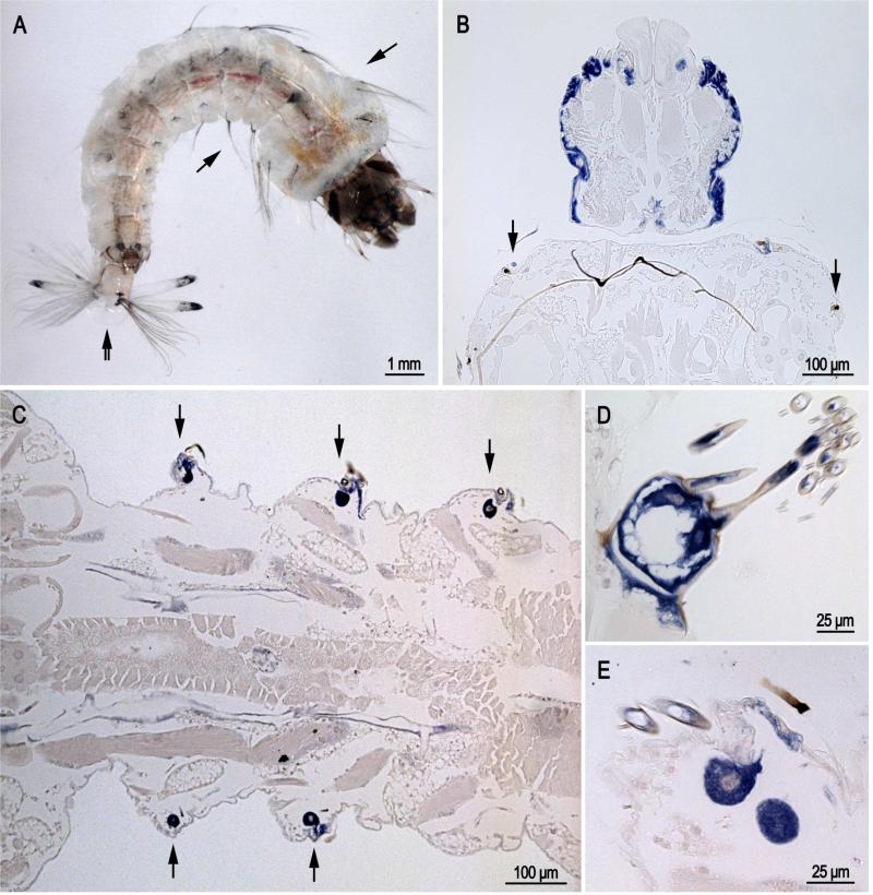
Fig. 2.
In situ hybridization of AgamCPCFC1 on sections of 4th instar larvae. A. Photograph of larva with arrows showing location of lateral setae on thorax and abdomen and a double arrow indicating the grid and fringe at the posterior end. B. Head capsule and bit of prothoracic segment. Note the presence of hybridization in the small cells that form setae at the anterior edge of the prothorax. C. Section of the abdomen showing cells that are forming setae. D. Grid and accompanying fringe at posterior end of a larva. E. Section showing cells secreting large and small setae. (B,D 3’ probe; C,E coding region probe).

